September 19, 2024
What To Anticipate During A Therapeutic Ultrasound
Ultrasound Physical Rehabilitation: Benefits, Side Effects & Treatments Certain, recommended healing acoustic wave are sent out deep in a constant or pulsating pattern right into the tissues when the transducer is revolved circularly over the injured location. Light heat is generated as the acoustic waves travel through the cells, increasing blood circulation in the location. The reduced strength ensures marginal heating, making it risk-free for more fragile or delicate locations while still experiencing the healing capability of ultrasound waves treatment. This ultrasound therapy strategy is frequently utilized in physical rehabilitation and rehabilitation setups to promote cells recovery, boost blood circulation, and ease discomfort. in various bone and joint conditions, such as pain in the back, arm joint injuries, knee sprains and even more. The primary objective of ultrasound treatment is to increase fast soft tissue recovery, boost blood circulation, and soothe pain in targeted areas. Phacoemulsification uses high-intensity ultrasound (1,000 W/cm2) simply put bursts (a couple of seconds) to piece and emulsify the lens during cataract surgical treatment.Cellular Results Of Ultrasound
Three months later on, an endothelial cell count was done, leading to Rejuvenation a substantial loss in the eyes subjected to phacoemulsification by ultrasound, along with in the corneal density. It was concluded that the femtosecond laser did not enhance the endothelial damages triggered by cataract surgical treatment, while using ultrasound did, hence showing that ultrasounds are unsafe for eyes with low endothelial cell values prior to undergoing surgical treatment. Ozil is a phacoemulsification cataract surgical procedure system established in 2005 in which ultrasonic waves with a regularity of 32 kHz were used continually or with bursts. This method achieves a boost in the efficiency of emulsification and a decrease in (or loss of) the repulsion in between the fragments generated.Ultrasound Of Bust Cyst
- Research studies by clinical associations and governmental bodies describe the recommended doses and kind of application for ultrasound utilized both physiotherapy and medication.
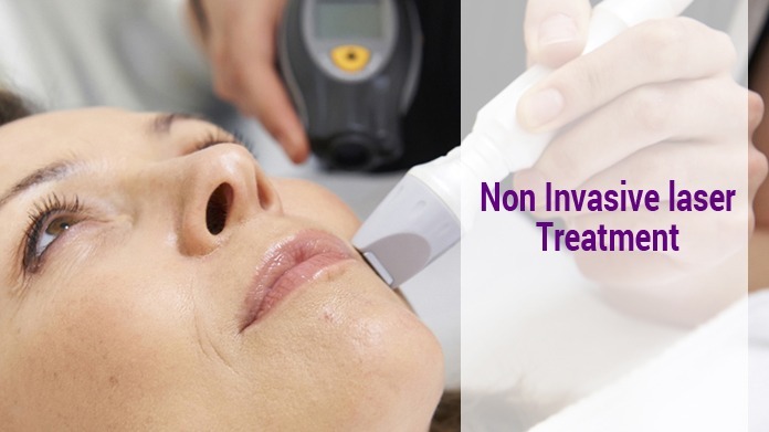
- This modality currently commonly has a base unit for producing an electric signal and a hand-held transducer.
- It is typical to keep these regularity restrictions for audible sound in between 20 Hz and 20 kHz, however these restrictions will certainly depend on the level of sensitivity of everyone [3]
- Low strength pulsed ultrasound has restorative application to speed up the recovery of bone cracks consisting of situations of nonunion (Gebauer et al. 2005).
What are the unfavorable negative effects of ultrasound therapy?
Ultrasound physical treatment has a reduced risk of triggering complications. But, exposure to low-intensity ultrasound for a very long time might trigger superficial burns on the skin. So, medical practitioners generally ensure that the ultrasound probe remains in movement when in contact with your skin.
Social Links